Using a new artificial intelligence identifying method, researchers at UC San Diego in La Jolla and their collaborators have taken what may turn out to be a leap forward in the understanding of human cells, according to a report published this week.
The pilot study — which combines microscopy, biochemistry techniques and artificial intelligence in a technique known as multi-scale integrated cell, or MuSIC — revealed about 70 components contained in a human kidney cell line, half of which had never been seen before.
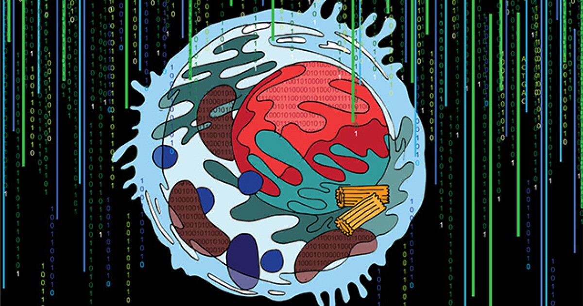
“If you imagine a cell, you probably picture the colorful diagram in your cell biology textbook, with mitochondria, endoplasmic reticulum and nucleus. But is that the whole story? Definitely not,” said Trey Ideker, a professor at UCSD’s School of Medicine and Moores Cancer Center. “Scientists have long realized there’s more that we don’t know than we know, but now we finally have a way to look deeper.”
The results were described in the Nov. 24 issue of Nature.
In one example, the researchers spotted a group of proteins forming an unfamiliar structure. Working with UCSD colleague Gene Yeo, they determined the structure to be a new complex of proteins that binds RNA. The complex likely is involved in splicing, a cellular event that enables the translation of genes to proteins and helps determine which genes are activated at which times.
The scientists had been interested in mapping the inner workings of cells for many years. What’s different about MuSIC is the use of deep learning to map the cell directly from cellular microscopy images.
“The combination of these technologies is unique and powerful because it’s the first time measurements at vastly different scales have been brought together,” said study first author Yue Qin, a bioinformatics and systems biology graduate student in Ideker’s lab.
Microscopes enable scientists to see to the level of a single micron — about the size of some organelles such as mitochondria. Smaller elements, such as individual proteins and protein complexes, can’t be seen through a microscope. Biochemistry techniques, which start with a single protein, allow scientists to get down to the nanometer scale, or one-billionth of a meter.
The team trained the MuSIC artificial intelligence platform to look at all the data and construct a model of the cell. The system doesn’t yet map the cell contents to specific locations, like a textbook diagram, partly because their locations aren’t necessarily fixed, the researchers said.
Ideker noted this was a pilot study to test MuSIC. The team has looked at only 661 proteins and one cell type.
“The clear next step is to blow through the entire human cell and then move to different cell types, people and species,” Ideker said. “Eventually we might be able to better understand the molecular basis of many diseases by comparing what’s different between healthy and diseased cells.” ◆
"that" - Google News
November 28, 2021 at 12:00AM
https://ift.tt/3cSl4nZ
UCSD finds technique that could boost mapping and understanding of cell interiors - La Jolla Light
"that" - Google News
https://ift.tt/3d8Dlvv


Tidak ada komentar:
Posting Komentar